

- Advertisement -
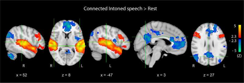
rest superimposed onto the MNI152 standard brain template. Brain regions activated during speech processing are shown in red-yellow, while deactivated in blue-green; p < 0.05, corrected with threshold-free cluster enhancement. Credit: NeuroImage (2026). DOI: 10.1016/j.neuroimage.2025.121638" width="800" height="438"/>
Activation maps resulting…
